OCT and Visual Field Testing
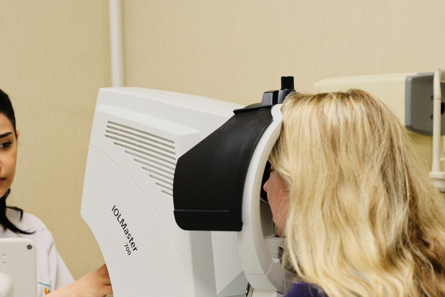
Measuring eye pressure alone is not sufficient for glaucoma follow-up.
Different tests are used to assess the structure of the optic nerve and changes over time. Among the most important are OCT imaging and visual field testing.
What Is OCT?
OCT (Optical Coherence Tomography) is an imaging method that provides detailed assessment of the optic nerve and retinal layers.
It allows structural changes associated with glaucoma to be assessed and results obtained during follow-up to be compared over time.
What Is a Visual Field Test?
A visual field test is a functional test that assesses peripheral vision.
It is used to monitor the effects of glaucoma on vision and identify possible changes.
Why Are OCT and Visual Field Tests Used Together?
OCT and visual field testing are complementary examinations.
One assesses the structure of the optic nerve, while the other provides information about changes in visual function.
They are therefore often assessed together during glaucoma follow-up.
Part of Regular Follow-Up
The aim in glaucoma is not only to assess the current condition, but also to monitor changes over time.
OCT and visual field tests are therefore important components of regular glaucoma follow-up.